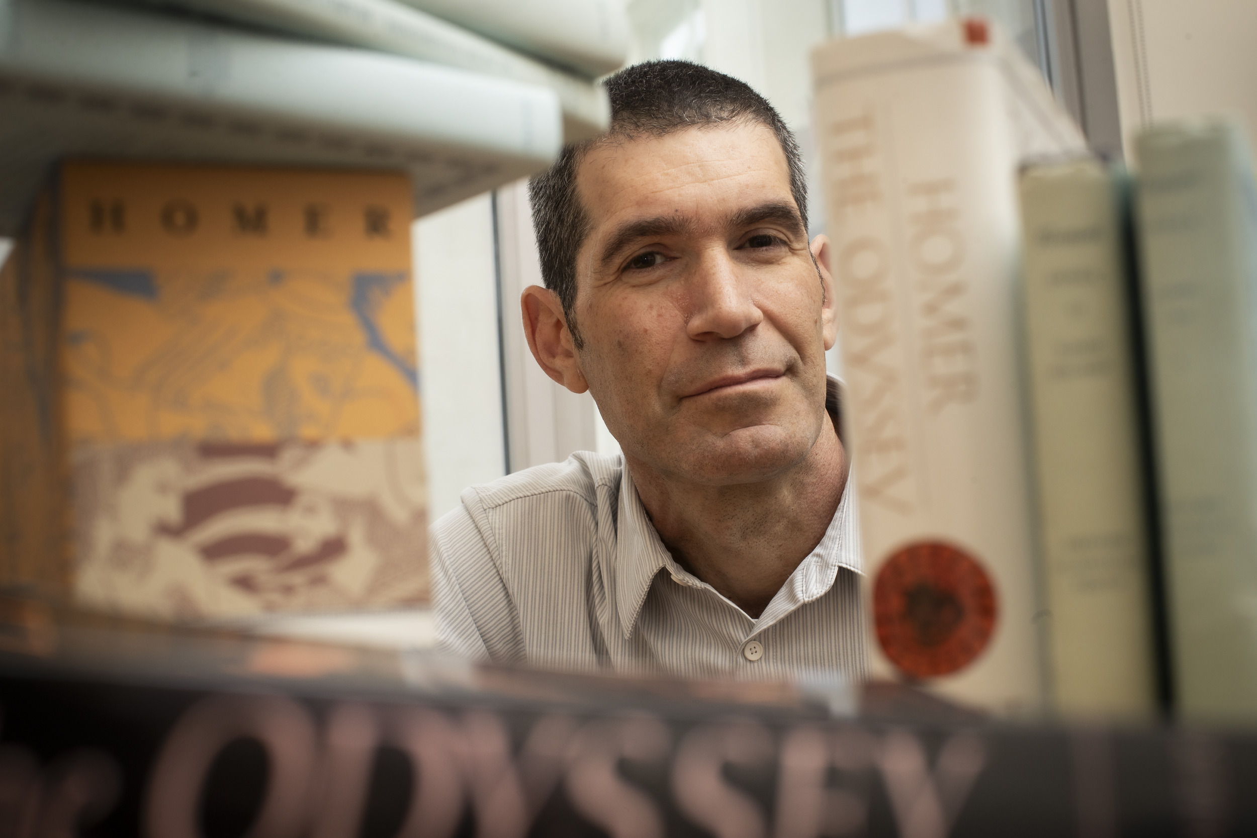
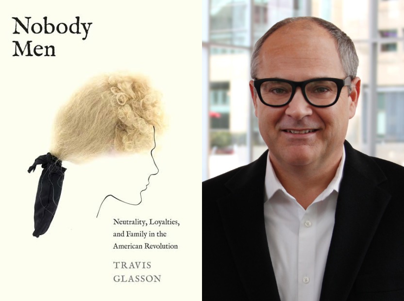
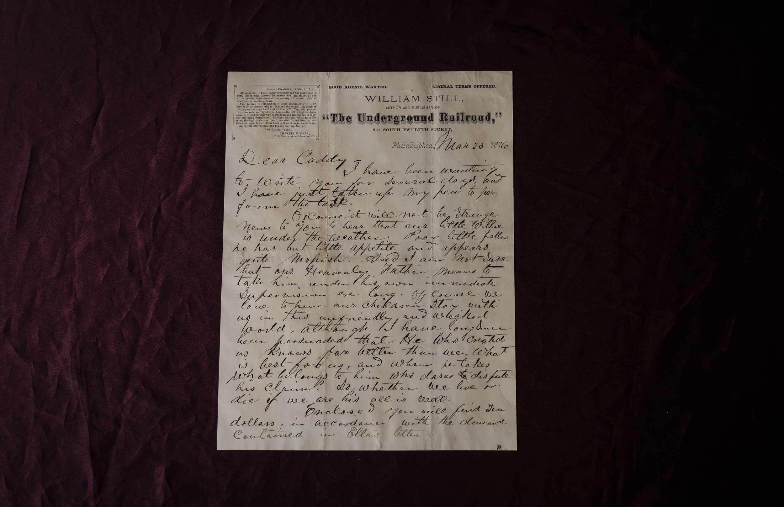
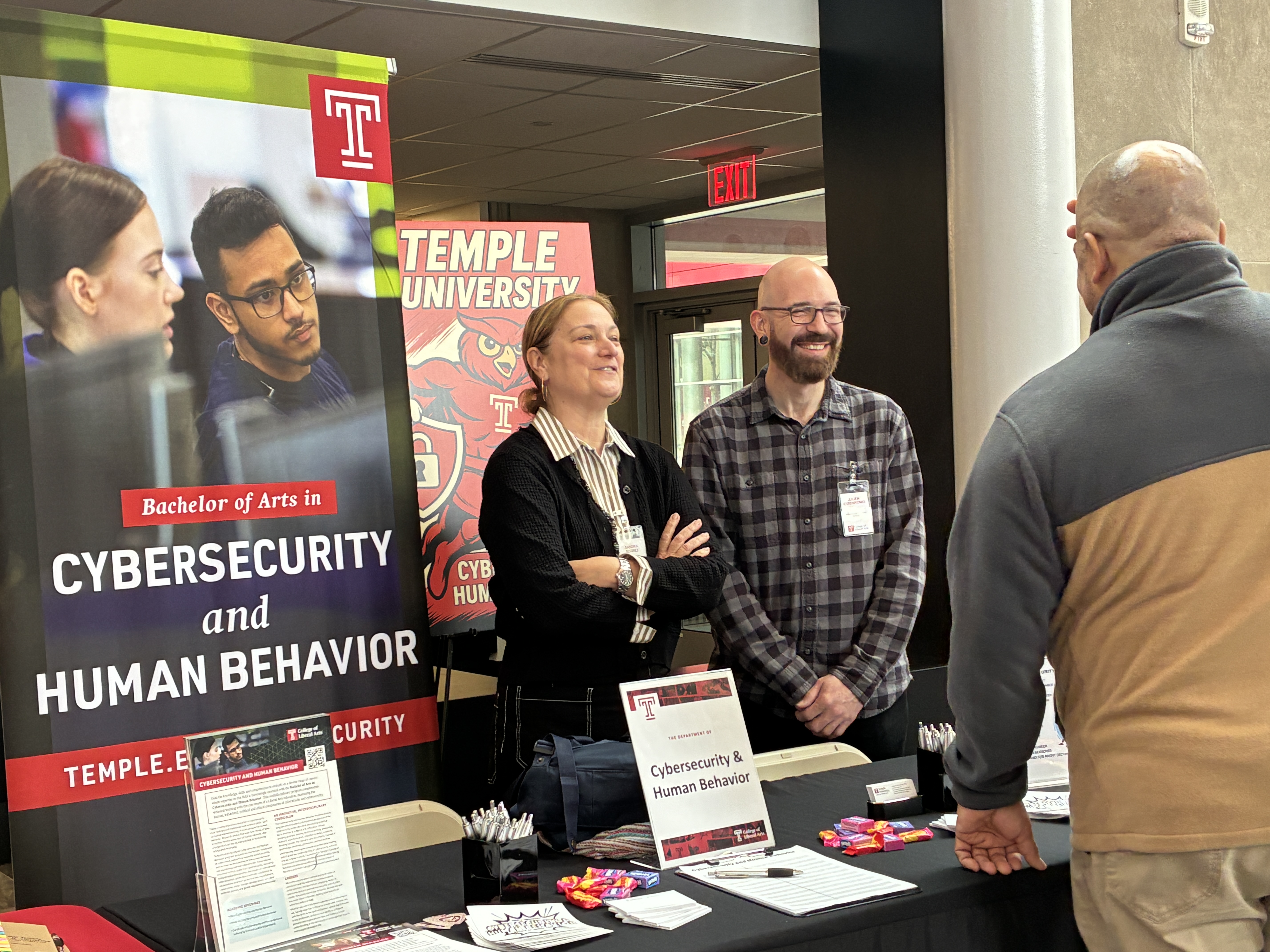
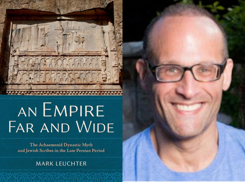
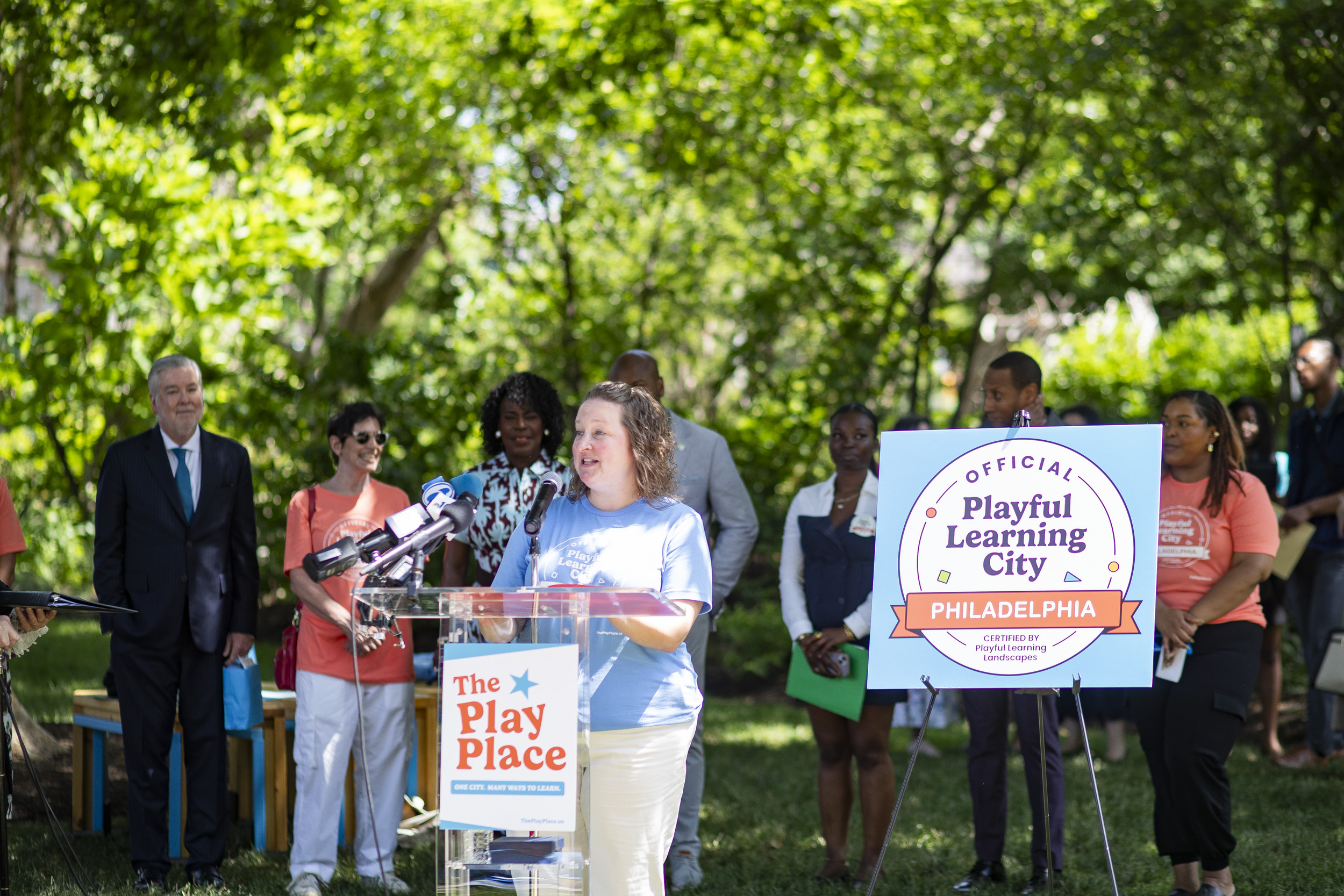
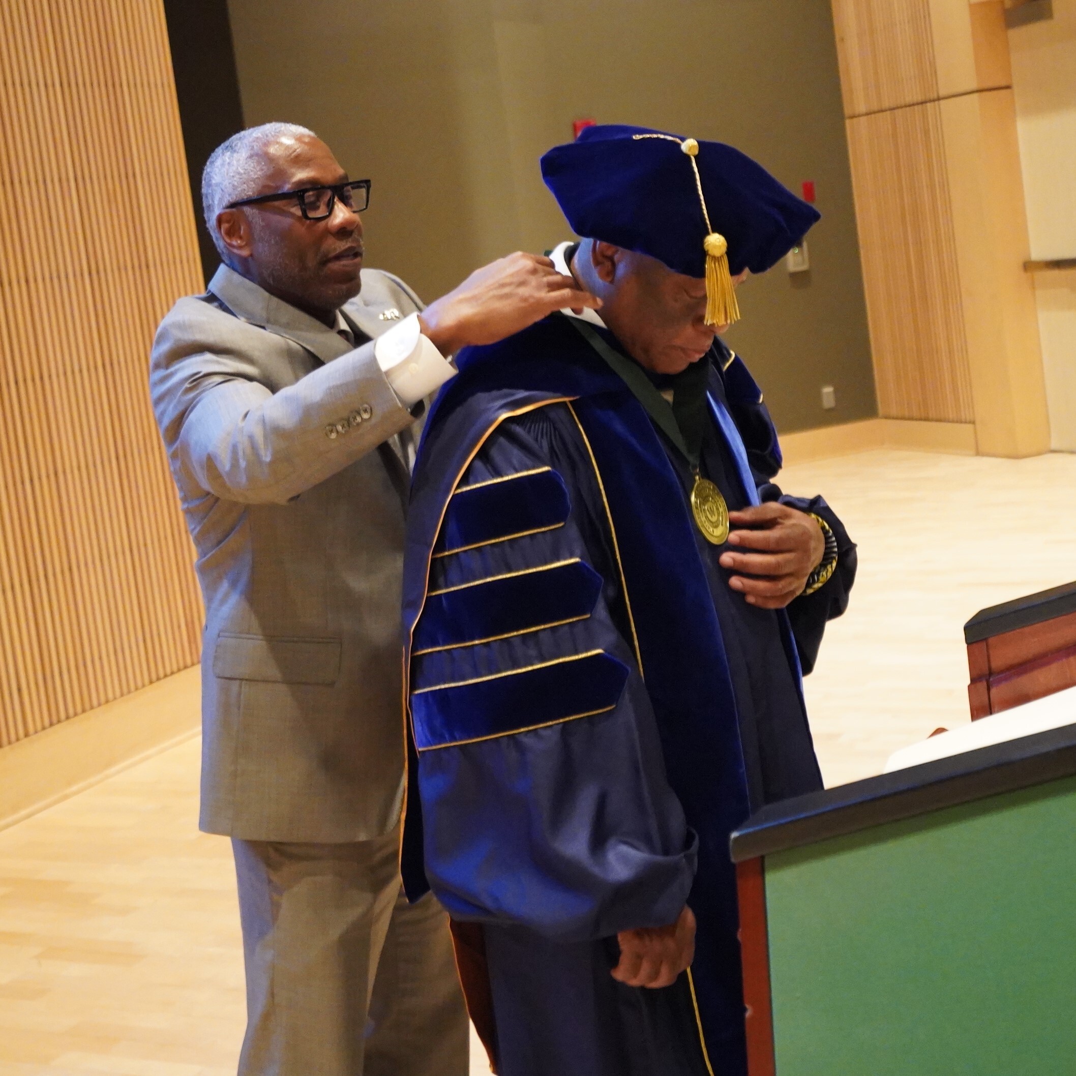
Jul. 16, 2026 Dr. Marisa Booty joins Criminal Justice Department faculty By Criminal Justice DepartmentThe Department of Criminal Justice is excited to welcome Dr. Marisa Booty to our faculty. Dr. Booty […] Jul. 9, 2026 Economics Department's Joshua Mask explores impact of AI tools on student outcomes in International Review of Economics Education By Economics DepartmentProfessor Joshua Mask has published a new paper titled Does AI Help Economics Students Learn […] Jul. 9, 2026 Economics Department's Viviane Sanfelice published in Labour Economics By Economics DepartmentProfessor Viviane Sanfelice has published a new paper titled "Public Childcare and Maternal […] Jul. 9, 2026 Economics Professor Michael Leeds receives Larry Hadley Award from NAASE By Economics DepartmentThe North American Association of Sports Economists presented Professor Peter von Allmen (Temple […] Photo by Ryan S. Brandenberg Jul. 8, 2026 Why we keep returning to ‘The Odyssey’ In anticipation of Christopher Nolan’s The Odyssey, set to be released in theaters on July 17, Dan […] Jul. 2, 2026 History professor Travis Glasson receives James Kirby Martin Book Prize By Jonathan HernandezThe Fort Plain Museum has awarded Travis Glasson, associate professor and chair of the History […] Photo by Betsy Manning Jun. 24, 2026 Temple researchers use GIS technology to uncover Philadelphia’s Underground Railroad history Using geographic information systems, College of Liberal Arts professors Jeremy Mennis and Nilgün […] Jun. 23, 2026 Inside the Cybersecurity and Human Behavior Program’s first year at CLA By Cybersecurity and Human Behavior ProgramIn the fall of 2025, the College of Liberal Arts introduced its new Cybersecurity and Human […] Jun. 15, 2026 Religion Professor Mark Leuchter receives Scott Award from Canadian Society of Biblical Studies By Jonathan HernandezThe College of Liberal Arts congratulates Professor of Religion Mark Leuchter on receiving an […] Photo by Ryan S. Bradenberg Jun. 15, 2026 Professor and former doctorate student team up to put research about play on the map By Matt PetrilloPhiladelphia was named the world’s first ‘Playful Learning […] Jun. 4, 2026 New book by Laura McGrath is reviewed in NYRB By Jonathan HernandezAssistant Professor of English Laura McGrath’s new book, Middlemen: Literary Agents and the Making […] Jun. 3, 2026 Talia LaSane receives NSF-funded Law and Science Dissertation Grant By Criminal Justice DepartmentCriminal Justice PhD candidate Talia LaSane has been awarded a highly competitive NSF-funded Law […] Toy Story 5 will be one of the first mainstream platforms to tackle the topic of AI and how it affects children, as the film will introduce a new character in Lilypad, a lightweight electronic tablet that competes for the affection of protagonist Bonnie. Temple Now caught up with Kathy Hirsh-Pasek, the Stanley and Debra Lefkowitz Faculty Fellow in the Department of Psychology, to gain her thoughts on the fictional animated film's relevance given the rising popularity of AI and electronic toys.Photo by Andrew Collette Jun. 3, 2026 ‘Toy Story 5’ to set stage for debate over AI in children’s toys Later this month, Toy Story 5 will introduce a new character with the electronic tablet Lilypad. […] Photo by Ryan S. Brandenberg May 15, 2026 Assistant professor of history named finalist for Pulitzer Bench Ansfield, assistant professor of history in the College of Liberal Arts, has been named a […] May 15, 2026 Molefi Kete-Asante awarded inaugural SUNY Old Westbury Presidential Medal By Jonathan HernandezThe State University of New York (SUNY) at Old Westbury has awarded Temple Africology and African […] Load More
Jul. 16, 2026 Dr. Marisa Booty joins Criminal Justice Department faculty By Criminal Justice DepartmentThe Department of Criminal Justice is excited to welcome Dr. Marisa Booty to our faculty. Dr. Booty […] Jul. 9, 2026 Economics Department's Joshua Mask explores impact of AI tools on student outcomes in International Review of Economics Education By Economics DepartmentProfessor Joshua Mask has published a new paper titled Does AI Help Economics Students Learn […] Jul. 9, 2026 Economics Department's Viviane Sanfelice published in Labour Economics By Economics DepartmentProfessor Viviane Sanfelice has published a new paper titled "Public Childcare and Maternal […] Jul. 9, 2026 Economics Professor Michael Leeds receives Larry Hadley Award from NAASE By Economics DepartmentThe North American Association of Sports Economists presented Professor Peter von Allmen (Temple […] Photo by Ryan S. Brandenberg Jul. 8, 2026 Why we keep returning to ‘The Odyssey’ In anticipation of Christopher Nolan’s The Odyssey, set to be released in theaters on July 17, Dan […] Jul. 2, 2026 History professor Travis Glasson receives James Kirby Martin Book Prize By Jonathan HernandezThe Fort Plain Museum has awarded Travis Glasson, associate professor and chair of the History […] Photo by Betsy Manning Jun. 24, 2026 Temple researchers use GIS technology to uncover Philadelphia’s Underground Railroad history Using geographic information systems, College of Liberal Arts professors Jeremy Mennis and Nilgün […] Jun. 23, 2026 Inside the Cybersecurity and Human Behavior Program’s first year at CLA By Cybersecurity and Human Behavior ProgramIn the fall of 2025, the College of Liberal Arts introduced its new Cybersecurity and Human […] Jun. 15, 2026 Religion Professor Mark Leuchter receives Scott Award from Canadian Society of Biblical Studies By Jonathan HernandezThe College of Liberal Arts congratulates Professor of Religion Mark Leuchter on receiving an […] Photo by Ryan S. Bradenberg Jun. 15, 2026 Professor and former doctorate student team up to put research about play on the map By Matt PetrilloPhiladelphia was named the world’s first ‘Playful Learning […] Jun. 4, 2026 New book by Laura McGrath is reviewed in NYRB By Jonathan HernandezAssistant Professor of English Laura McGrath’s new book, Middlemen: Literary Agents and the Making […] Jun. 3, 2026 Talia LaSane receives NSF-funded Law and Science Dissertation Grant By Criminal Justice DepartmentCriminal Justice PhD candidate Talia LaSane has been awarded a highly competitive NSF-funded Law […] Toy Story 5 will be one of the first mainstream platforms to tackle the topic of AI and how it affects children, as the film will introduce a new character in Lilypad, a lightweight electronic tablet that competes for the affection of protagonist Bonnie. Temple Now caught up with Kathy Hirsh-Pasek, the Stanley and Debra Lefkowitz Faculty Fellow in the Department of Psychology, to gain her thoughts on the fictional animated film's relevance given the rising popularity of AI and electronic toys.Photo by Andrew Collette Jun. 3, 2026 ‘Toy Story 5’ to set stage for debate over AI in children’s toys Later this month, Toy Story 5 will introduce a new character with the electronic tablet Lilypad. […] Photo by Ryan S. Brandenberg May 15, 2026 Assistant professor of history named finalist for Pulitzer Bench Ansfield, assistant professor of history in the College of Liberal Arts, has been named a […] May 15, 2026 Molefi Kete-Asante awarded inaugural SUNY Old Westbury Presidential Medal By Jonathan HernandezThe State University of New York (SUNY) at Old Westbury has awarded Temple Africology and African […]