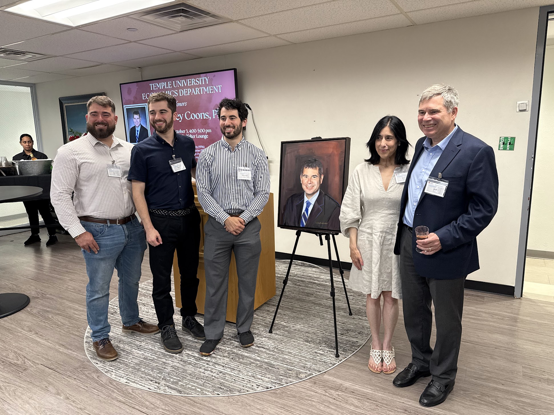
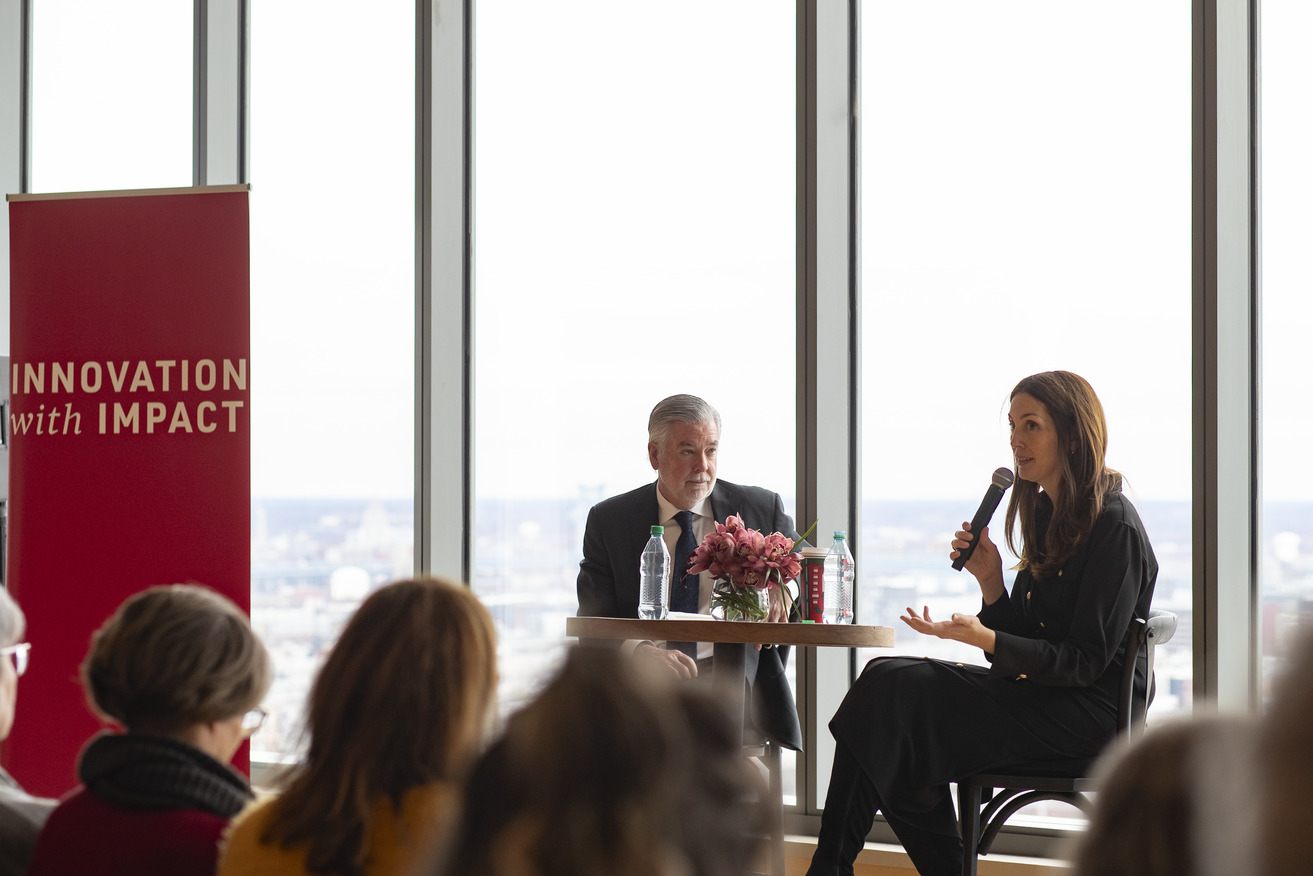
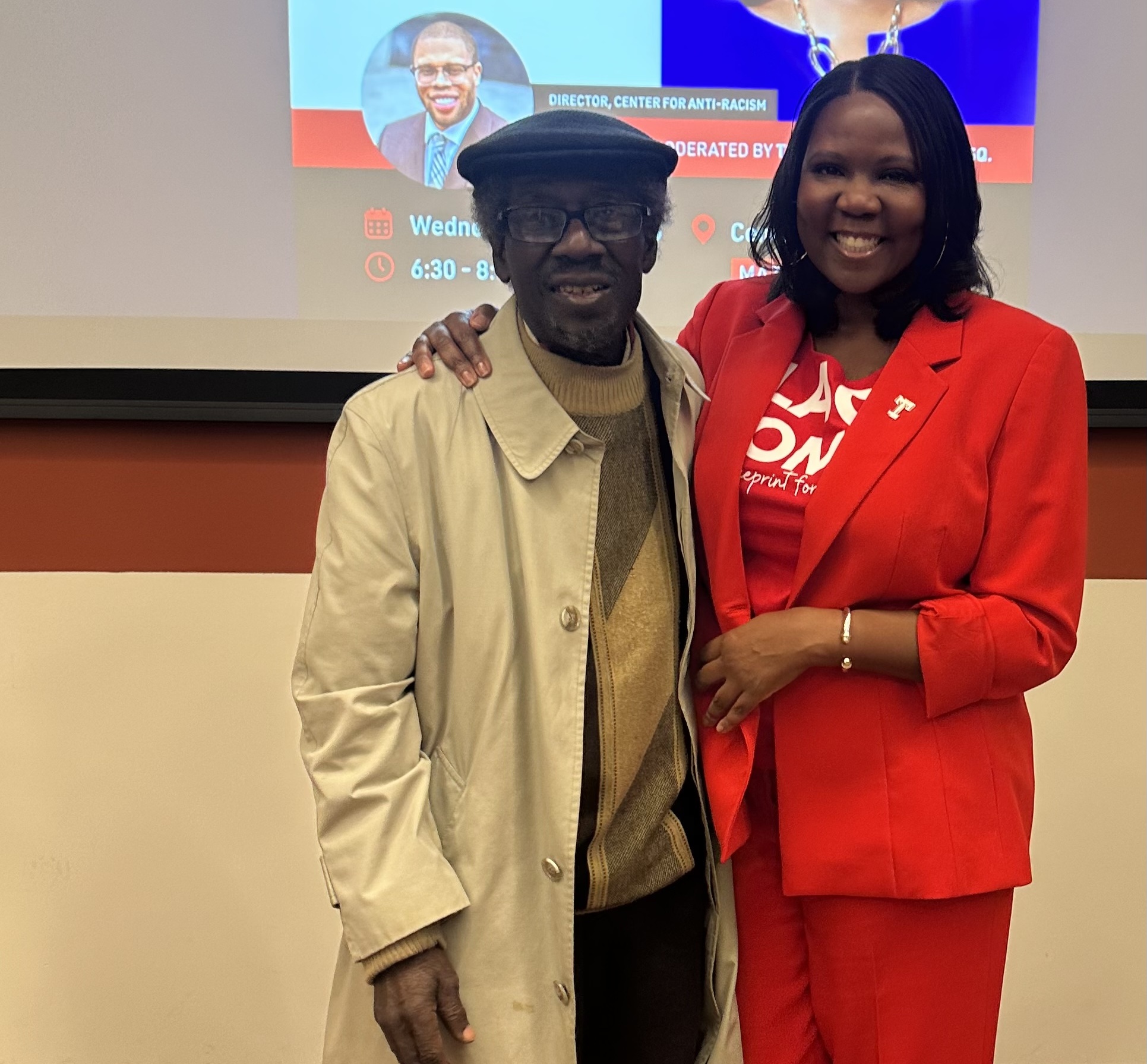
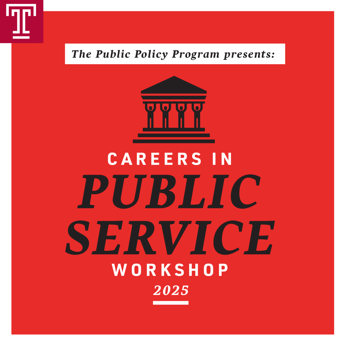
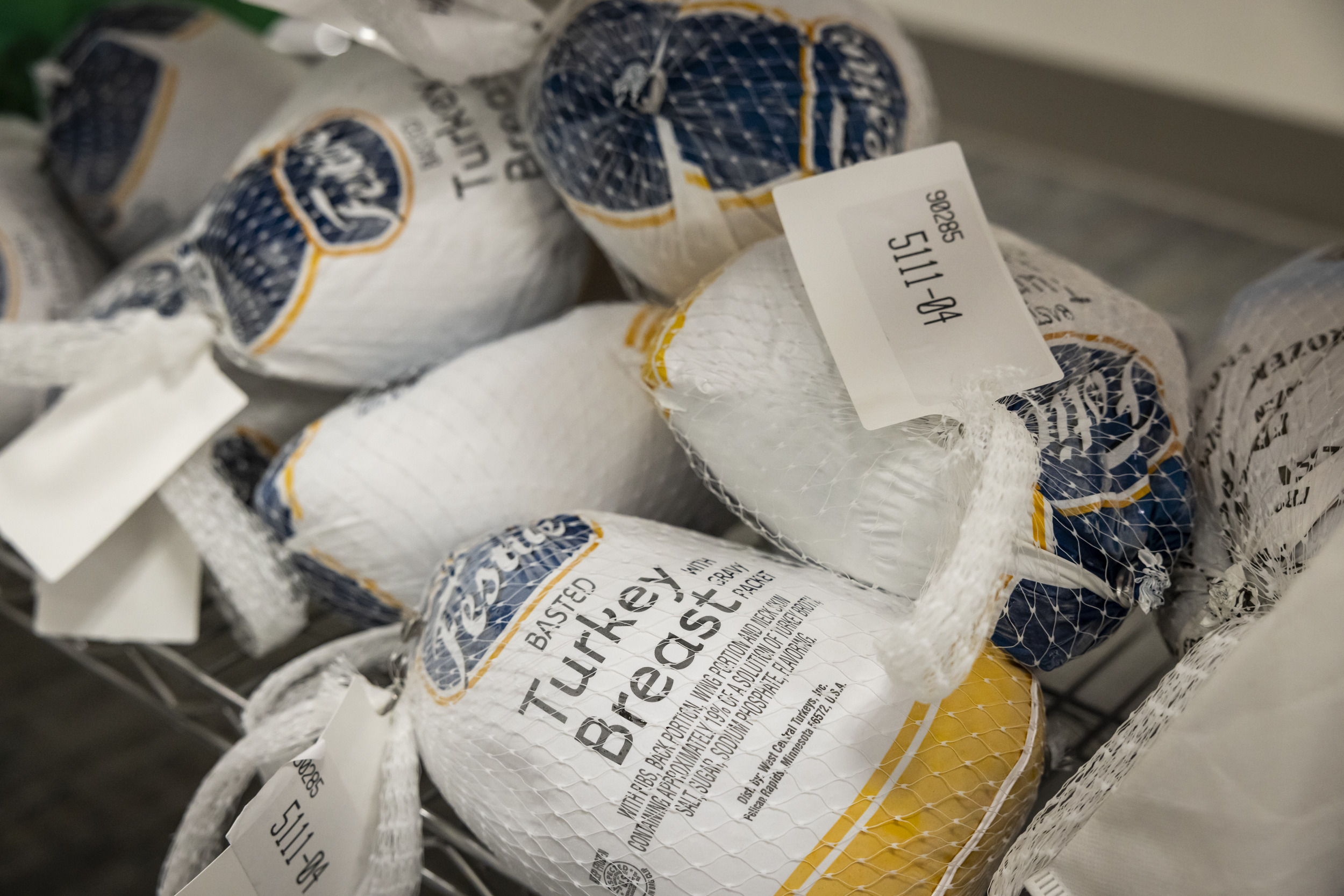
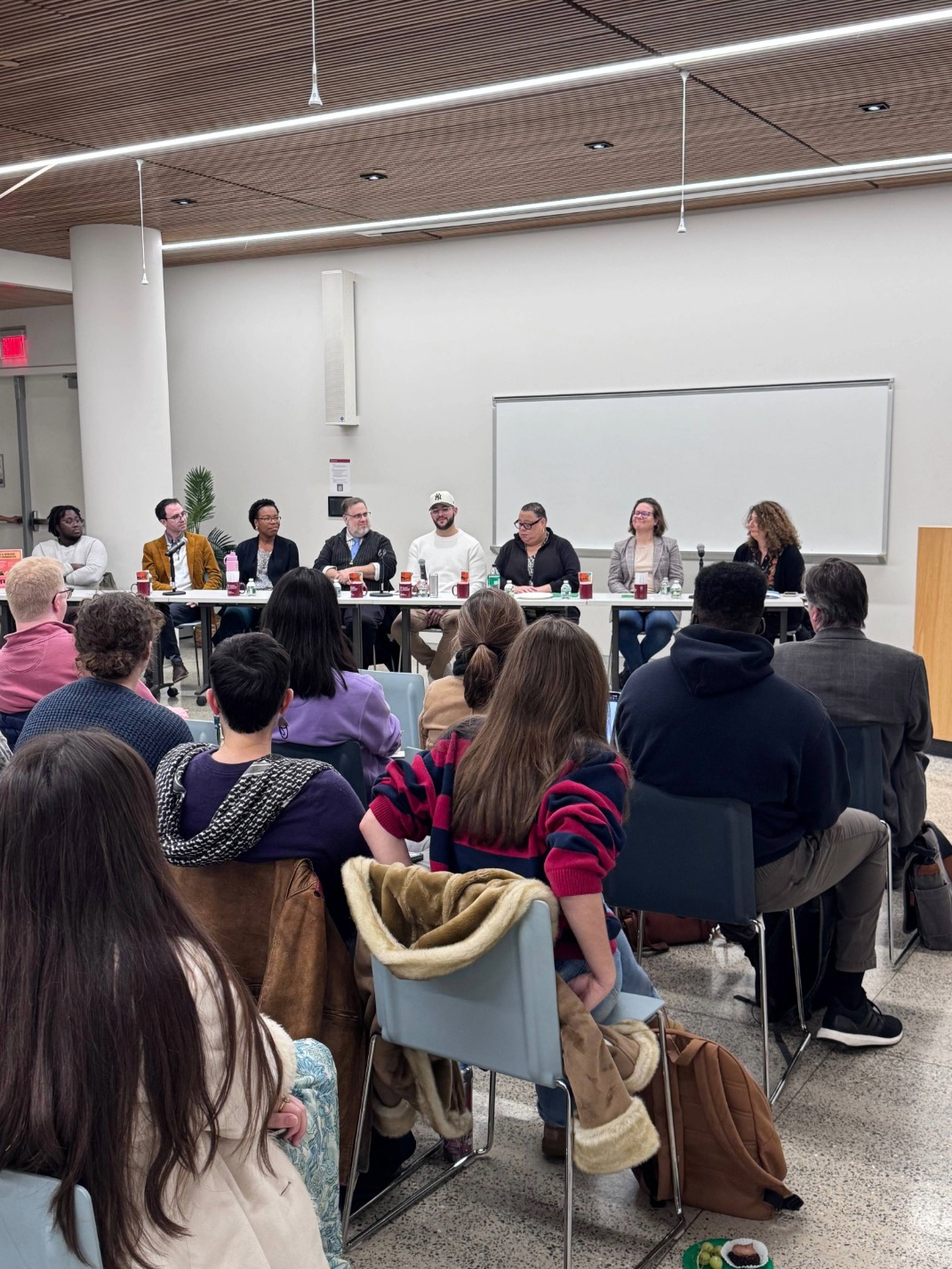
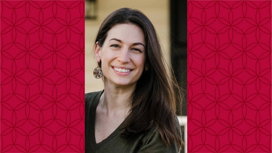
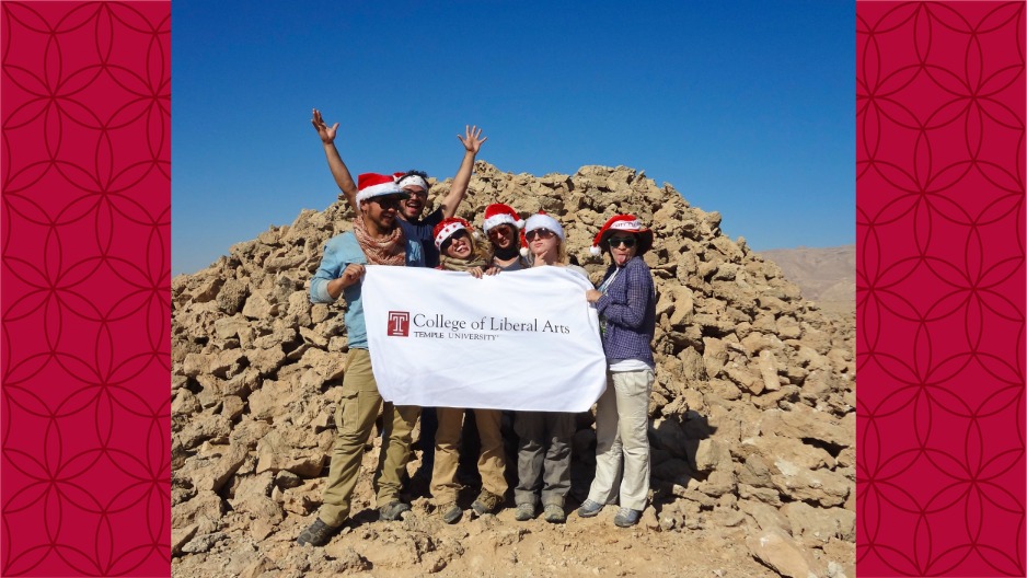
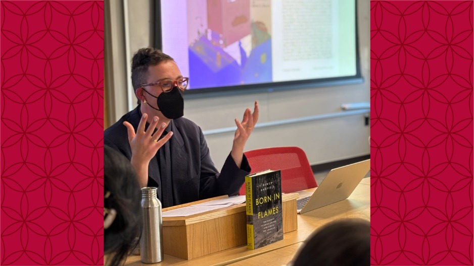
Jan. 6, 2026 Laura Levitt guest co-edits special issue of MAVCOR Journal By Jonathan HernandezProfessor of Religion Laura Levitt is credited as a guest co-editor in a special issue of MAVCOR […] Dec. 16, 2025 Sociology major wins grand prize at new Temple pitch competition By Jonathan HernandezFor young children, conversation is a powerful catalyst of emotional intelligence, self-expression […] The launch of Temple’s Innovation with Impact series featured a conversation between President John Fry and Professor Liz Moore about the intersections of storytelling, scholarship and community.Photo by Ryan S. Brandenberg Dec. 11, 2025 Temple’s Innovation with Impact series opens with conversation featuring Liz Moore The professor of English, director of the MFA program in creative […] Dec. 9, 2025 London summer program invites students to examine gender through film and theater By Jonathan HernandezThis summer, Temple students will have the opportunity to study in one of the world’s most dynamic […] Dec. 5, 2025 Internship Spotlight: Jasmine Mehta By Jonathan HernandezJasmine Mehta is a junior political science and philosophy major with a minor in public policy. […] Adjoa Asamoah is pictured with Emeritus Professor Abu Abarry during her recent visit back to Temple. Abarry served as one of her professors when she previously studied at the university.Photo by Timothy Welbeck Dec. 4, 2025 'Everything that I am' is because of Temple Two-time Temple alum Adjoa Asamoah, CLA ’98, EDU ’01, recently visited her alma mater where she […] Dec. 1, 2025 Temple’s Public Policy Program hosts the 2025 careers in public service workshop By Kasey TrappOn Saturday, November 15th, 2025, Temple’s Public Policy Program hosted its fifth Careers in Public […] Nov. 26, 2025 Africology and African American Studies Department celebrates Reynaldo Anderson’s book release By Jonathan HernandezOn October 22, the Charles L. Blockson Collection in Sullivan Hall hosted a celebration of Reynaldo […] According to Joshua Mask of the Economics Department in the College of Liberal Arts, Philadelphians will have to pay more for their turkeys than the rest of the country this year.Photo by Betsy Manning Nov. 24, 2025 Gobblenomics: Your turkey may be cheaper this year, but not if you live in Philadelphia It was just last month that it was reported that inflation had reached its highest level since this […] Panelists mulling over a question about how they personally weigh risks and rewards of public humanities work. From L to R: Tangie Wilson, Dan Blank, Heather Lewis-Weber, Michael Brix, Oscar Almonte-Espinal, Jamie Brunson, Elizabeth Kimball, Laurie Zierer. Nov. 20, 2025 Panel of local community leaders tackle risks and rewards of public humanities By Christina BakerDoing work in the humanities means telling stories and creating narratives about the communities […] Nov. 20, 2025 Alumni Spotlight: Neuroscience grad Sophie Farley continues developing professional pathways By Jonathan HernandezWhen Sophie Farley, CLA ‘12, arrived at Temple University, she didn’t yet know exactly where her […] Kim Williams poses with students in front of an uncovered tomb in Oman Nov. 7, 2025 Anthropology Professor Digs a Little Deeper For Student Success By Jonathan HernandezLong before she ever found herself excavating 5,000-year-old tombs in the deserts of Oman, Temple […] Nov. 4, 2025 PhD student Bertram Liyanage publishes a chapter in National Epics project By Religion DepartmentCongratulations to 3rd year PhD student, Bertram Liyanage, who was assigned to write the Sri Lanka […] Nov. 4, 2025 Economics major Renee Oymann maintains an edge in fencing and academics By Jonathan HernandezFencing, the swordfighting sport often called “physical chess,” demands a great deal of focus, […] Oct. 28, 2025 CHAT and History Department celebrate release of Bench Ansfield's new book By Center for the HumanitiesIn their monograph Born in Flames, Bench Ansfield, Assistant Professor of History, examines the […] Load More
Jan. 6, 2026 Laura Levitt guest co-edits special issue of MAVCOR Journal By Jonathan HernandezProfessor of Religion Laura Levitt is credited as a guest co-editor in a special issue of MAVCOR […] Dec. 16, 2025 Sociology major wins grand prize at new Temple pitch competition By Jonathan HernandezFor young children, conversation is a powerful catalyst of emotional intelligence, self-expression […] The launch of Temple’s Innovation with Impact series featured a conversation between President John Fry and Professor Liz Moore about the intersections of storytelling, scholarship and community.Photo by Ryan S. Brandenberg Dec. 11, 2025 Temple’s Innovation with Impact series opens with conversation featuring Liz Moore The professor of English, director of the MFA program in creative […] Dec. 9, 2025 London summer program invites students to examine gender through film and theater By Jonathan HernandezThis summer, Temple students will have the opportunity to study in one of the world’s most dynamic […] Dec. 5, 2025 Internship Spotlight: Jasmine Mehta By Jonathan HernandezJasmine Mehta is a junior political science and philosophy major with a minor in public policy. […] Adjoa Asamoah is pictured with Emeritus Professor Abu Abarry during her recent visit back to Temple. Abarry served as one of her professors when she previously studied at the university.Photo by Timothy Welbeck Dec. 4, 2025 'Everything that I am' is because of Temple Two-time Temple alum Adjoa Asamoah, CLA ’98, EDU ’01, recently visited her alma mater where she […] Dec. 1, 2025 Temple’s Public Policy Program hosts the 2025 careers in public service workshop By Kasey TrappOn Saturday, November 15th, 2025, Temple’s Public Policy Program hosted its fifth Careers in Public […] Nov. 26, 2025 Africology and African American Studies Department celebrates Reynaldo Anderson’s book release By Jonathan HernandezOn October 22, the Charles L. Blockson Collection in Sullivan Hall hosted a celebration of Reynaldo […] According to Joshua Mask of the Economics Department in the College of Liberal Arts, Philadelphians will have to pay more for their turkeys than the rest of the country this year.Photo by Betsy Manning Nov. 24, 2025 Gobblenomics: Your turkey may be cheaper this year, but not if you live in Philadelphia It was just last month that it was reported that inflation had reached its highest level since this […] Panelists mulling over a question about how they personally weigh risks and rewards of public humanities work. From L to R: Tangie Wilson, Dan Blank, Heather Lewis-Weber, Michael Brix, Oscar Almonte-Espinal, Jamie Brunson, Elizabeth Kimball, Laurie Zierer. Nov. 20, 2025 Panel of local community leaders tackle risks and rewards of public humanities By Christina BakerDoing work in the humanities means telling stories and creating narratives about the communities […] Nov. 20, 2025 Alumni Spotlight: Neuroscience grad Sophie Farley continues developing professional pathways By Jonathan HernandezWhen Sophie Farley, CLA ‘12, arrived at Temple University, she didn’t yet know exactly where her […] Kim Williams poses with students in front of an uncovered tomb in Oman Nov. 7, 2025 Anthropology Professor Digs a Little Deeper For Student Success By Jonathan HernandezLong before she ever found herself excavating 5,000-year-old tombs in the deserts of Oman, Temple […] Nov. 4, 2025 PhD student Bertram Liyanage publishes a chapter in National Epics project By Religion DepartmentCongratulations to 3rd year PhD student, Bertram Liyanage, who was assigned to write the Sri Lanka […] Nov. 4, 2025 Economics major Renee Oymann maintains an edge in fencing and academics By Jonathan HernandezFencing, the swordfighting sport often called “physical chess,” demands a great deal of focus, […] Oct. 28, 2025 CHAT and History Department celebrate release of Bench Ansfield's new book By Center for the HumanitiesIn their monograph Born in Flames, Bench Ansfield, Assistant Professor of History, examines the […]